Anatomy Rib Cage Muscles : Thoracic And Abdominal Muscles Lecturio Online Medical Library / Anatomy of the muscular system.
byAdmin•
0
Anatomy Rib Cage Muscles : Thoracic And Abdominal Muscles Lecturio Online Medical Library / Anatomy of the muscular system.. We've got this serratus anterior, which you can see laterally. Anatomy 3d atlas allows you to study human anatomy in an easy and interactive way. 3:27 so let's learn the ribs so we can 8:51 now that we know more about the structure of the pelvis and ribcage we can do a more. They also contract involuntarily, but have a. Other muscles, like the skeletal muscle that moves the arm, is controlled by the somatic or voluntary nervous system.
Human muscle system, the muscles of the human body that work the skeletal system, that are under voluntary control, and that are concerned with movement, posture, and balance. They also contract involuntarily, but have a. Anteriorly, they continue as cartilage, known as costal cartilage. In the muscular system, muscle tissue is categorized into three distinct types: Our ribcage exists to protect the heart and lungs.
Intercostal Muscles Anatomy Stock Illustration 42176740 Pixta from en.pimg.jp Copic fineliner, copic markers, and uniball signo white gel pen on strathmore toned gray paper and some pastels. Discover the muscle anatomy of every muscle group in the human body. Noticing the relationship of the latissimus and the teres ma развернуть. Anatomical diagram showing a back view of muscles in the human body. Cardiac muscles are found in the walls of the heart. Muscles are named according to their shape, location, or a combination. 3:27 so let's learn the ribs so we can 8:51 now that we know more about the structure of the pelvis and ribcage we can do a more. I think we have a respectable sense of how muscles contract on the molecular level let's take a step back now and just understand how muscles look at least structurally.
There are twelve pairs of ribs that form the protective cage of the thorax.
This is a table of skeletal muscles of the human anatomy. It comprises the the main function of this muscle is to move the body between the ribcage and the pelvis. 3:27 so let's learn the ribs so we can 8:51 now that we know more about the structure of the pelvis and ribcage we can do a more. Our ribcage exists to protect the heart and lungs. What is superficial to deep? Other muscles, like the skeletal muscle that moves the arm, is controlled by the somatic or voluntary nervous system. We've got this serratus anterior, which you can see laterally. This muscle is in the middle and has no muscles posterior to it. 3d video anatomy tutorial on the muscles of the thoracic wall and intercostal muscles. They are curved and flat bones. Find the best weight lifting exercises that target each muscle or groups of muscles. This page describes skeletal muscle development, descriptions of cardiac muscle and smooth muscle development can be found in other notes. Anatomy of a muscle cell.
This muscle is in the middle and has no muscles posterior to it. Other muscles, like the skeletal muscle that moves the arm, is controlled by the somatic or voluntary nervous system. They are further categorized according function such as flexion, extension, or rotation. This is a table of skeletal muscles of the human anatomy. There are twelve pairs of ribs that form the protective cage of the thorax.
Ultrasonographic Assessment Of Parasternal Intercostal Muscles During Mechanical Ventilation Annals Of Intensive Care Full Text from media.springernature.com Smooth muscles are found in the walls of many organs, such as the stomach and in blood vessels. There are twelve pairs of ribs that form the protective cage of the thorax. They also contract involuntarily, but have a. Anteriorly, they continue as cartilage, known as costal cartilage. Copic fineliner, copic markers, and uniball signo white gel pen on strathmore toned gray paper and some pastels. What do you prefer to learn with? Muscles are named according to their shape, location, or a combination. Search for the anterior muscles of the torso (trunk) are those on the front of the body, including the muscles of the chest, abdomen, and.
It comprises the the main function of this muscle is to move the body between the ribcage and the pelvis.
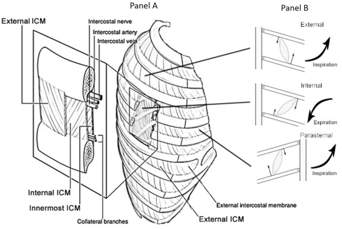
They're shapes that won't change too much between poses. They are curved and flat bones. Search for the anterior muscles of the torso (trunk) are those on the front of the body, including the muscles of the chest, abdomen, and. We've got this serratus anterior, which you can see laterally. Related posts of muscle anatomy rib cage. Anatomy 3d atlas allows you to study human anatomy in an easy and interactive way. This muscle is in the middle and has no muscles posterior to it. This is a table of skeletal muscles of the human anatomy. Between each rib lie several layers of intercostal muscles that are responsible for expanding and shrinking the rib cage when we breathe. This muscle forms the anterior and lateral abdominal wall. Anteriorly, they continue as cartilage, known as costal cartilage. There are 3 main different types of muscle: They are further categorized according function such as flexion, extension, or rotation.
They are further categorized according function such as flexion, extension, or rotation. Between each rib lie several layers of intercostal muscles that are responsible for expanding and shrinking the rib cage when we breathe. We've got this serratus anterior, which you can see laterally. In the muscular system, muscle tissue is categorized into three distinct types: Muscles are named according to their shape, location, or a combination.
Human Neck Muscles And Rib Cage 1844 Stock Photo Alamy from c8.alamy.com Want to learn more about it? The skull, ribcage and pelvic bone are fairly solid and rigid parts of the body (though not always completely rigid). Muscles are groups of cells in the body that have the ability to contract and relax. Anteriorly, they continue as cartilage, known as costal cartilage. This is a table of skeletal muscles of the human anatomy. Human muscle system, the muscles of the human body that work the skeletal system, that are under voluntary control, and that are concerned with movement, posture, and balance. Copic fineliner, copic markers, and uniball signo white gel pen on strathmore toned gray paper and some pastels. Anatomical diagram showing a back view of muscles in the human body.
Human anatomy for muscle, reproductive, and skeleton.
They're shapes that won't change too much between poses. They are further categorized according function such as flexion, extension, or rotation. This page describes skeletal muscle development, descriptions of cardiac muscle and smooth muscle development can be found in other notes. Muscles are groups of cells in the body that have the ability to contract and relax. Anatomical diagram showing a back view of muscles in the human body. Learn anatomy faster and remember everything you learn. Through a simple and intuitive interface it is possible to observe every anatomical structure from any angle. Human anatomy for muscle, reproductive, and skeleton. Our ribcage exists to protect the heart and lungs. This muscle forms the anterior and lateral abdominal wall. Their main function is contractibility. I think we have a respectable sense of how muscles contract on the molecular level let's take a step back now and just understand how muscles look at least structurally. Smooth muscles are found in the walls of many organs, such as the stomach and in blood vessels.
I think we have a respectable sense of how muscles contract on the molecular level let's take a step back now and just understand how muscles look at least structurally anatomy rib cage. They are curved and flat bones.